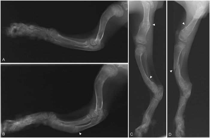
Figure 2.
Radiographic images of the forelimbs and hindlimbs of a kitten with severe osteogenesis imperfecta. A. Lateral view of the right forelimb. B. Lateral view of the left forelimb. The radius and ulna are curved with respect to the right. A fracture can be seen in the ulna (arrowhead). C. Lateral view of the right hindlimb. D. Lateral view of the left hindlimb. There are bilateral femoral and tibial fractures (arrowheads).

