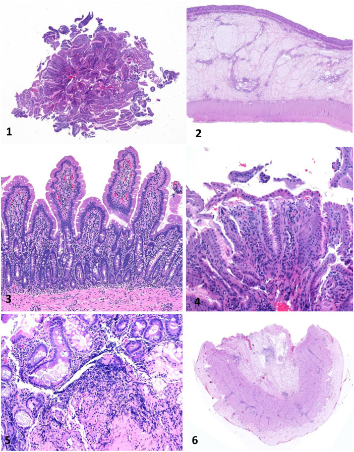
Figures 1–6.
Equine gastrointestinal biopsies. Figure 1. Gastric endoscopic biopsy showing only mucosa sectioned tangentially. H&E. Figure 2. Colon surgical biopsy, including full thickness of the colonic wall. H&E. Figure 3. Duodenal surgical biopsy. This biopsy is mostly free of artifact and well oriented, so that numerous villus-crypt units with associated lamina propria can be evaluated. H&E. Figure 4. Gastric endoscopic biopsy that includes only the superficial aspect of the mucosa with artifactually detached mucosal epithelium. H&E. Figure 5. Duodenal endoscopic biopsy with severe crushing artifact. H&E. Figure 6. Duodenal biopsy with muscularis and serosa, but missing mucosa and submucosa. H&E.

