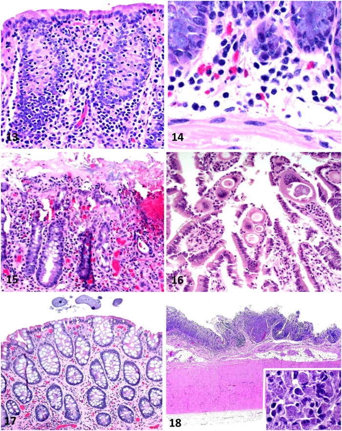
Figures 13–18.
Equine intestinal biopsies. Figure 13. Loss of goblet cells in the colonic mucosa of a horse with chronic diarrhea. This is a nonspecific lesion in the colon of horses with diarrhea. H&E. Figure 14. Small numbers of eosinophils in the deep lamina propria of the small intestine. This is considered a normal background finding in horses and should not be interpreted as a significant lesion. H&E. Figure 15. Eosinophilic proctitis; large numbers of eosinophils are intermixed with macrophages, lymphocytes, and plasma cells within the lamina propria. The cause of this lesion was not determined. H&E. Figure 16. Eimeria leuckarti in the lamina propria of the small intestine; this parasite is usually considered an incidental finding in horses. H&E. Figure 17. Ciliated protozoa in the lumen of the colon; these parasites are usually considered an incidental finding in horses. H&E. Figure 18. Granulomatous colitis in a horse with Rhodococcus equi infection. Macrophage infiltrates are present. Inset: small coccobacilli are present in the cytoplasm of most macrophages H&E.

