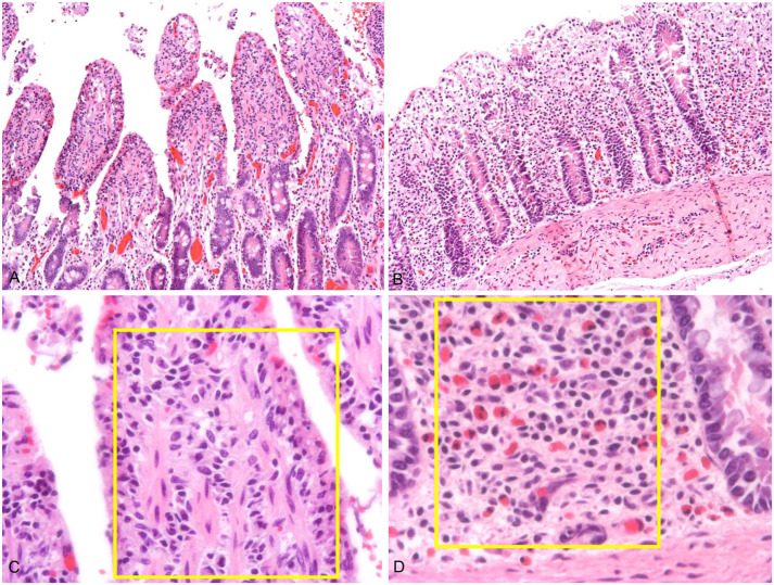
Figure 4.
Representative images of small and large intestinal mucosa, with mild autolysis. A. Low magnification of jejunal mucosa. H&E. B. Low magnification of left ventral colon. H&E. C. High magnification of the jejunal villus lamina propria. The yellow rectangle is an area of interest. H&E. D. High magnification of the deep lamina propria of the left ventral colon. The yellow rectangle is the area of interest. Note the higher number of eosinophils than in the jejunal deep lamina propria. H&E.

