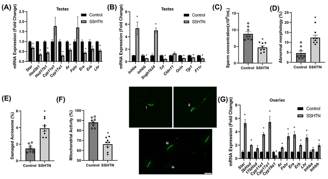
Figure 3. SSHTN mice exhibited gonadal dysfunction.

Gene expression of steroidogenic pathway genes and hormone receptors in (A) testes from control and mice-administered L-NAME in the drinking water for 2 weeks, then 2 weeks of washout, and then a subsequent 3 weeks of 4% salt diet (SSHTN), n=6. Gene expression of secretory proteins and tight junction proteins in (B) testes from control and SSHTN mice, n=6. Sperm functional tests in control and SSHTN mice demonstrate (C) decreased sperm concentration, n=8, (D) increased percentage of sperm with abnormal morphology, n=8, (E) increased percentage of sperm with damaged acrosome, n=8, and (F) increased number of sperm with nonfunctional mitochondrial activity n=8. Representative images of (i) intact and (ii) damaged acrosome integrity were assessed using FITC-PNA that binds exclusively to the outer membrane of the acrosome. Scale bars = 10 μm. Florescent dye Rh123 (Rhodamine123) distinguishes (iii) functional and (iv) nonfunctional sperm mitochondria as only live cells can retain the stain after washing. Scale bars = 20 μm. Expression of steroidogenic pathway genes, hormone receptors, and secretory proteins in (G) ovaries from control and SSHTN mice, n=6. Results are expressed as mean ± SEM, and statistical analysis consisted of a Student’s t test. *P<0.05 vs control.
