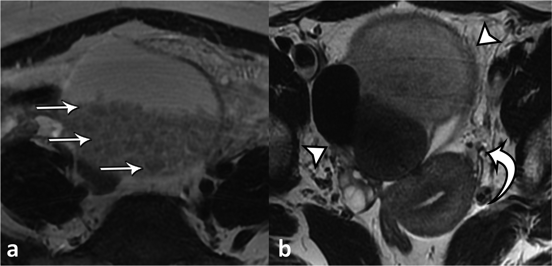
Figure 2.

a) Axial T2-weighted MRI of the pelvis demonstrating large mixed cystic and solid mass centered in the midline pelvis with numerous rounded globules (straight arrows) are seen within, reflecting the “boba sign”. The cyst contents exhibited iso- to hypointense signal on T1-weighted imaging and did not show signal drop-out on out-of-phase imaging or fat suppression or internal enhancement was observed (not shown). b) Axial T2-weighted MRI of the pelvis shows a large, multilocular mass with components with iso- and hypointense signal (arrowheads). The uterus is tilted towards the left (curved arrow), indicative of torsion. Engorged gonadal vessels were also present (not shown).
