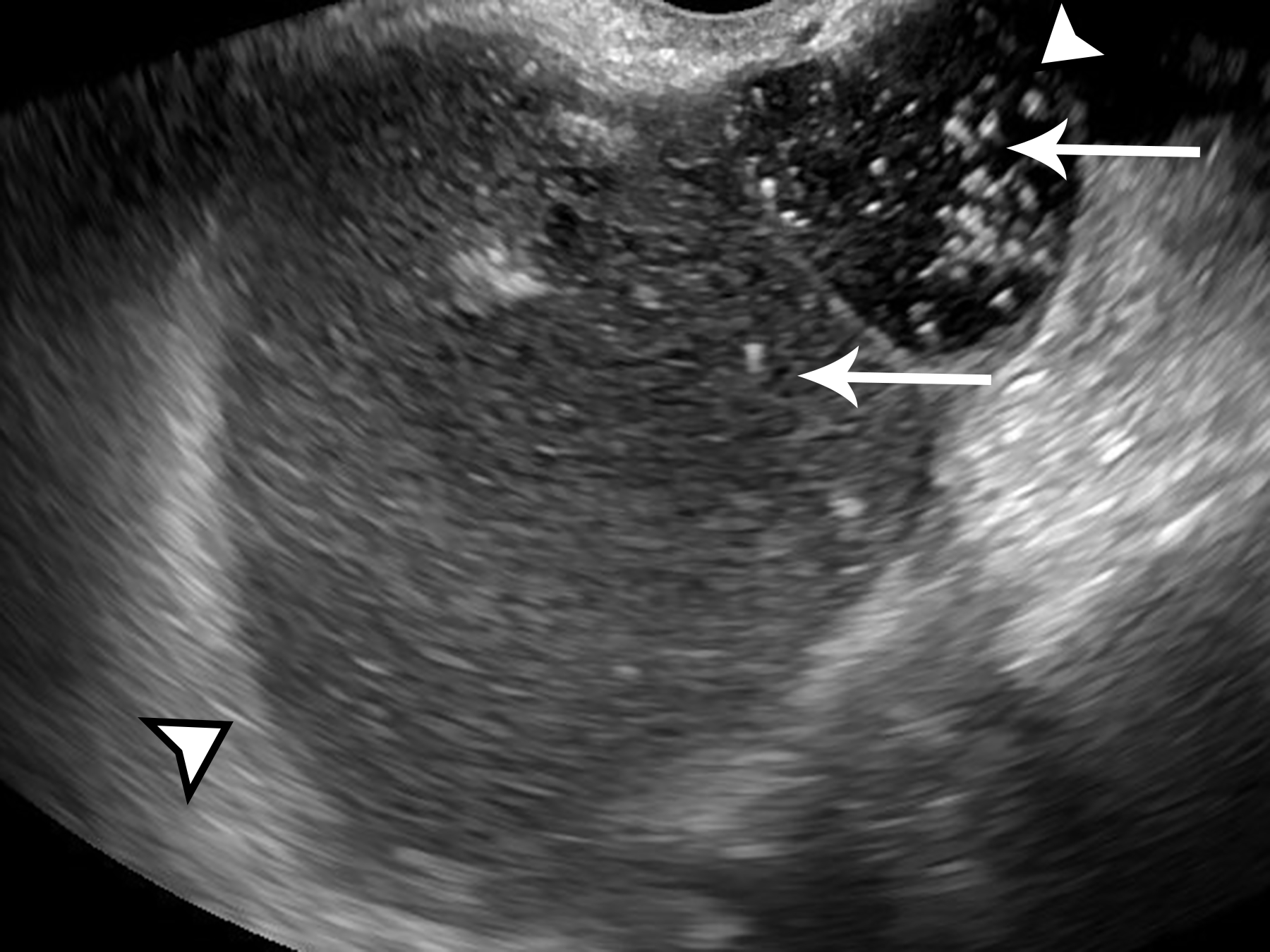
Figure 3.

Ultrasound image of the left adnexa in the longitudinal plane, obtained with endovaginal technique, shows multilocular cystic mass with varying internal echogenicity (arrowheads). Multiple echogenic foci with comet-tail artifact are present (arrows).
