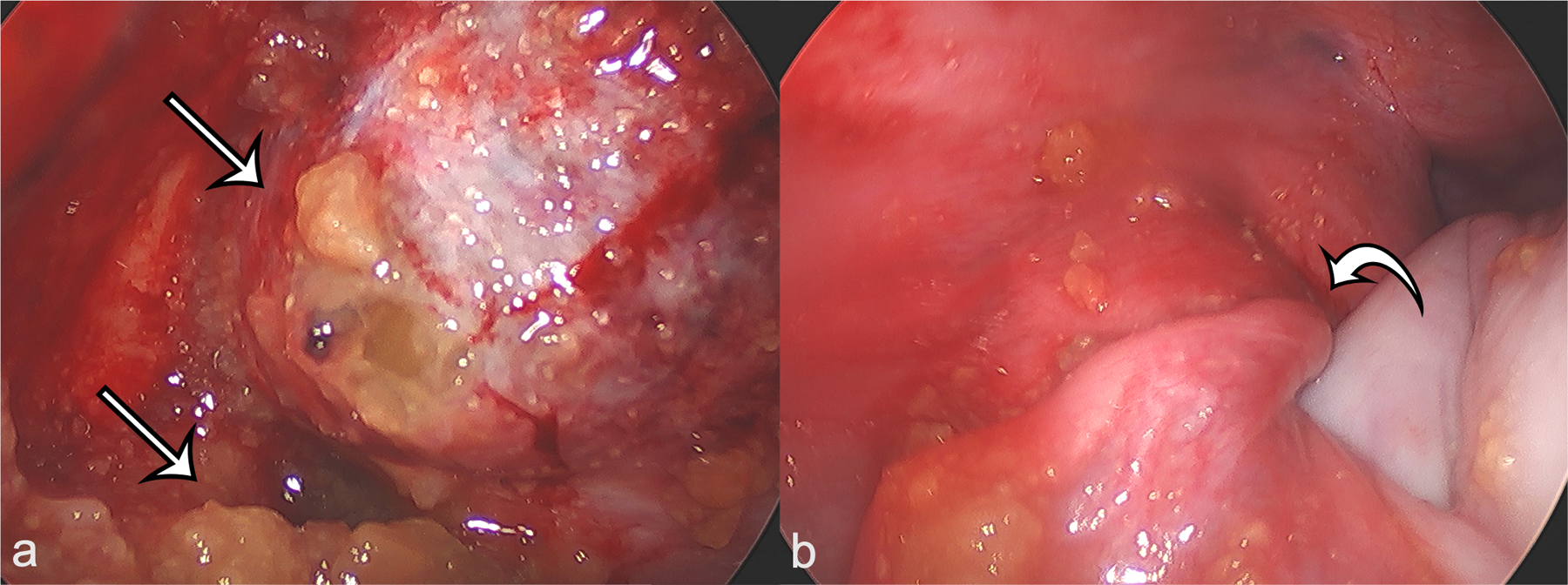
Figure 4.

a) Intraoperative laparoscopic image of the left adnexa demonstrates ruptured yellow, gelatinous contents (arrows) of the teratoma, which were confirmed to be thyroid colloid on microscopic review. b) Intraoperative laparoscopic image of the left adnexa demonstrates twisting of the left infundibulopelvic ligament (curved arrow), confirming the diagnosis of ovarian torsion.
