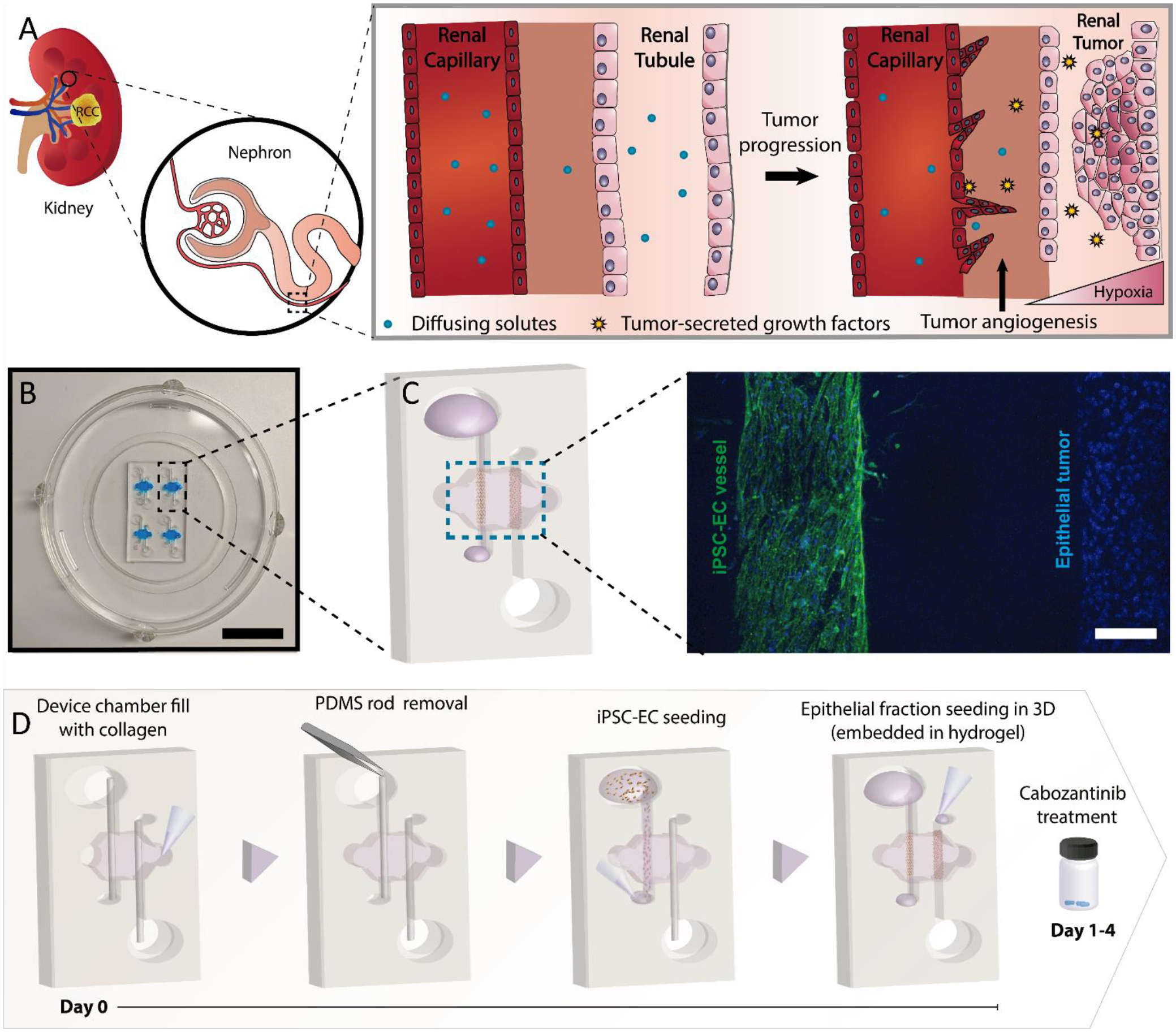
Figure 1: Renal cell carcinoma on a chip.

Schematic illustrating the biological processes reproduced in the microdevice. A) Illustration of the tumor progression and angiogenesis model. The model includes a renal capillary and epithelial tubule as a renal tumor develops, along with the increase in angiogenesis in the nearby vasculature. B) Picture of the microdevice and schematic of the co-culture organization in the device. C) Projected confocal image of an iEC-Epithelial co-culture stained with CD31 (green, iEC) and DAPI (blue, all nuclei). D) Schematic depicting the co-culture procedure in the microdevice and timeline for the cabozantinib treatment. Cells shown on the right are epithelial cells.
