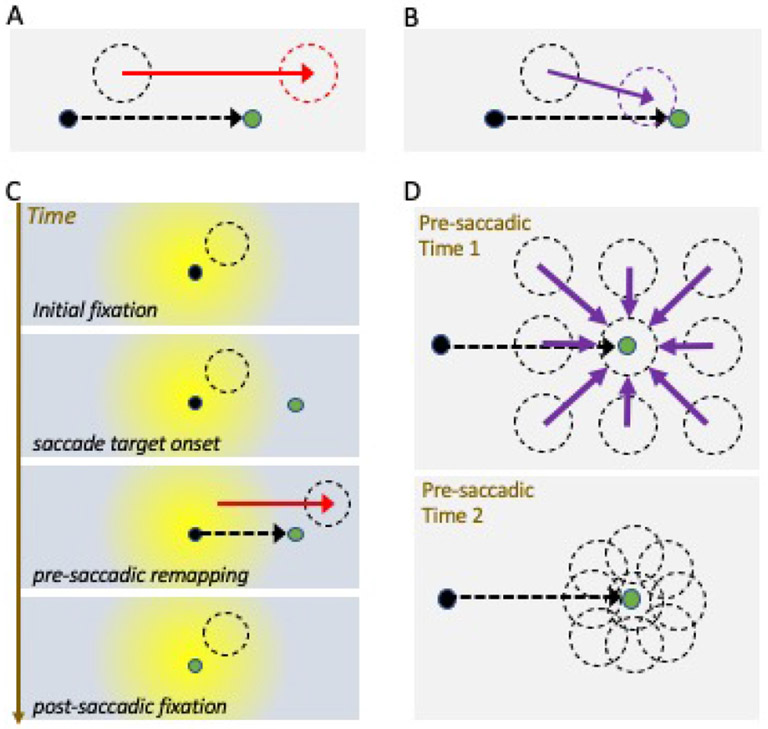
Figure 2: Forward vs convergent remapping.
(A-B) Single neuron schematic for forward spatial remapping (A) and convergent remapping (B). Black dot is initial fixation target (current gaze position), and green dot is saccade target. Black dashed circle depicts the neuron’s current receptive field (RF), red dashed circle indicates the forward remapping field location (future field), and purple indicates the convergent remapping field location (toward saccade target). (C) Time course of forward spatial remapping from a retinal perspective, with schematic centered on current gaze position at each time point. (Note Panel A depicts remapping instead from a world-centered perspective.) Timeframes illustrate how the retinal RF position changes under the influence of the saccade plan, shifting to a new retinal location during pre-saccadic remapping, and returning to the classical RF position once fixation is acquired at the saccade target (green). Yellow shading indicates the fovea and parafoveal area of the retina. (D) Convergent spatial remapping can enhance processing at saccade endpoints. All RFs shift towards the saccade target during remapping, resulting in an increase in the density of the neural representation around the saccade target that can mimic attentional gain effects.

