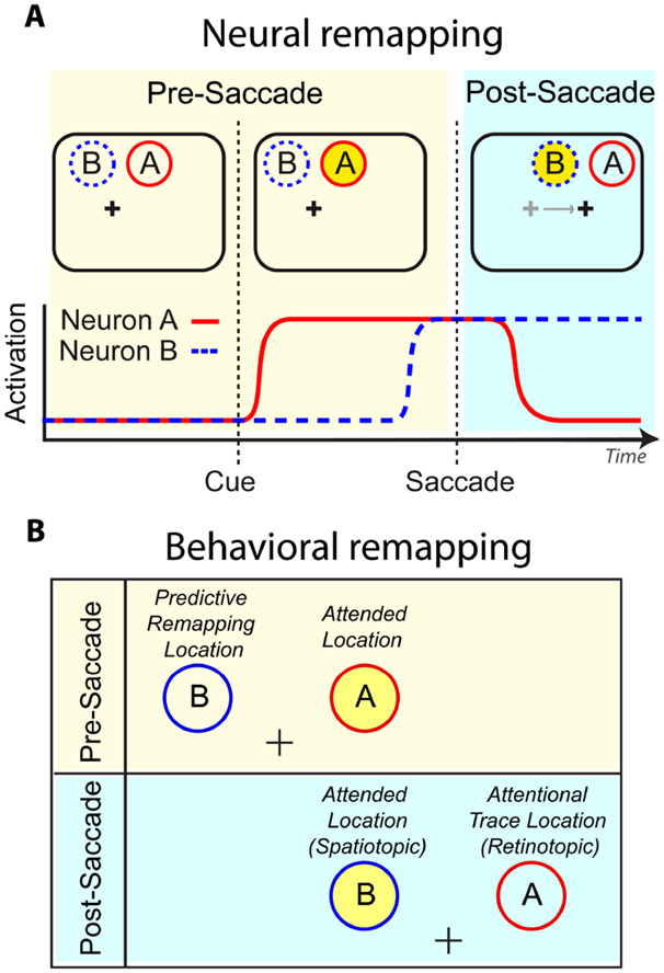
Figure 3. Predictive Remapping vs Retinotopic Attentional Trace.
(A) Hypothetical responses of two visual neurons with different spatial receptive fields. The beige interval indicates the period prior to saccade initiation, the blue interval after. Yellow circle represents to-be-attended spatiotopic location. Before the saccade, the attended location falls within Neuron A’s receptive field; after the saccade, it falls in Neuron B’s. ‘Predictive remapping’ is when Neuron B begins to respond in anticipation of the saccade. ‘Retinotopic attentional trace’ is when Neuron A continues to respond for a period of time after the eye movement. Thus, there is a period of time where both spatiotopic and retinotopic locations are facilitated. (B) Corresponding locations for a behavioral study. Figure adapted from Golomb 2019. (Note to Annual Reviews: I am an author of this article, and the publisher has confirmed that I retain the rights to re-publish it.)

