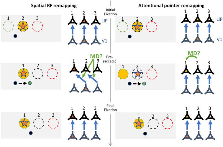
Figure 4. Spatial receptive field remapping vs attentional pointer remapping.
Left side: Schematic illustrating remapping mechanism where spatial receptive fields (RFs) transiently shift in anticipation of the saccade. Right side: Schematic illustrating alternative attentional pointer remapping mechanism. Gray boxes illustrate the locations of 3 neurons’ RFs (dashed circles), with current gaze position at the black dot, and the green dot representing the saccade target. The orange star indicates the stimulus probe, and the yellow shading indicates the attentional focus. Next to each box is a simplified diagram of the same 3 neurons (conceptualized here as LIP neurons), and the corresponding V1 neurons feeding feed-forward input (blue arrows indicate these connections). During the initial fixation period (top row), the stimulus falls in the RF of neuron 2. After the saccade (bottom row), the stimulus falls in the RF of neuron 1. During remapping (middle row), neuron 1 becomes active in anticipation of the saccade. The spatial RF remapping mechanism says this is because the RFs shift spatially to their future fields, which could be conceptualized as a remapping of which retinotopic V1 neurons feed into the LIP neurons, such that the neurons become transiently sensitive to a different portion of the visual field. The attentional pointer mechanism instead says that the RFs remain veridical, but the new set of neurons becomes facilitated in anticipation (i.e., the attention pointer remaps from neuron 2 to neuron 1). In both cases, the remapping signal could come from corollary discharge signals from an area such as thalamic MD.

