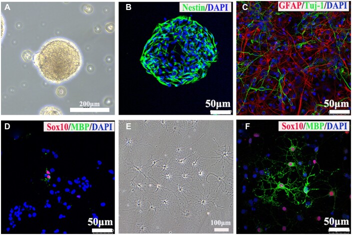
Figure 1.
Identifications of NSCs and OLGs. (A) The primary cultured NSCs formed neurospheres under light microscope. (B) Immunostaining image showed NSCs positively expressed nestin protein. (C, D) Images of immunostaining against tuj-1 (neuron marker), GFAP (astrocyte marker), SOX10 (OLGs marker) and MBP (mature OLGs marker) antibodies. (E) The primary mature OLGs extended multiple processes under light microscope. (F) Images of immunostaining against markers of OLGs including MBP and Sox10.

