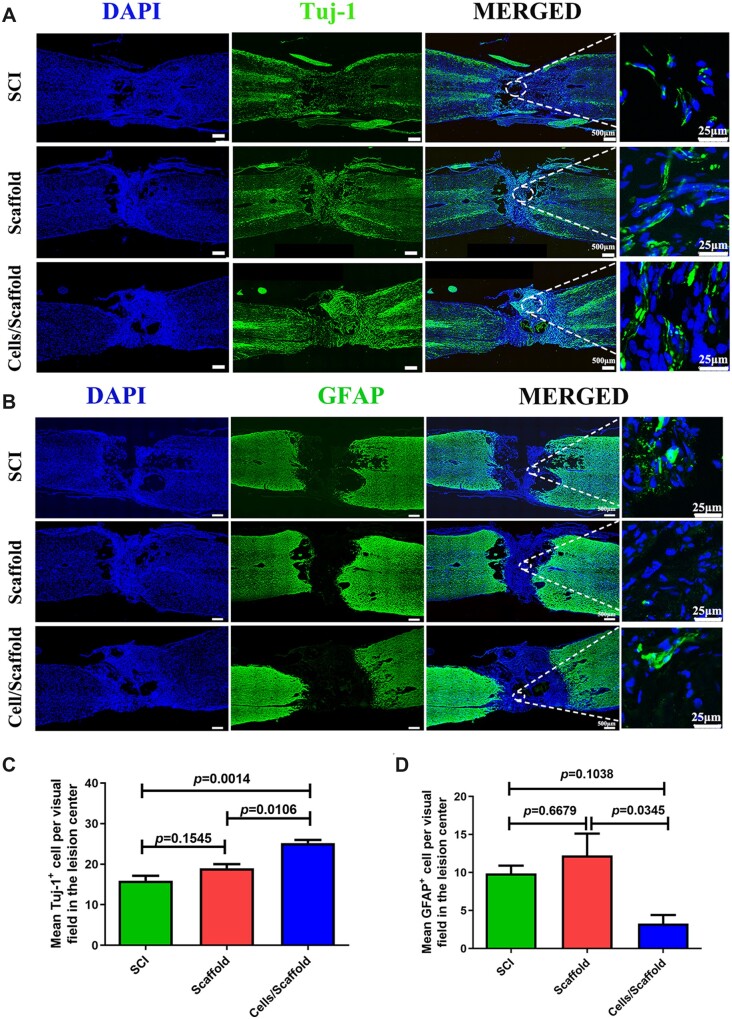
Figure 4.
Three-dimensional bioprinting scaffold with NSCs and OLGs promoted nerve regeneration and inhibited astrocytic formation in vivo. (A, B) Immunofluorescence stainings for tuj-1 and GFAP. A much larger number of tuj-1-positive cells were observed in the cells/scaffold group compared with the scaffold and SCI groups. The quantity of GFAP-positive astrocytes at lesion site area in the cells/scaffold group was lower than that in the SCI and scaffold groups. (C, D) Quantification of tuj-1-positive cells and GFAP-positive cells in SCI, scaffold and cells/scaffold groups.

