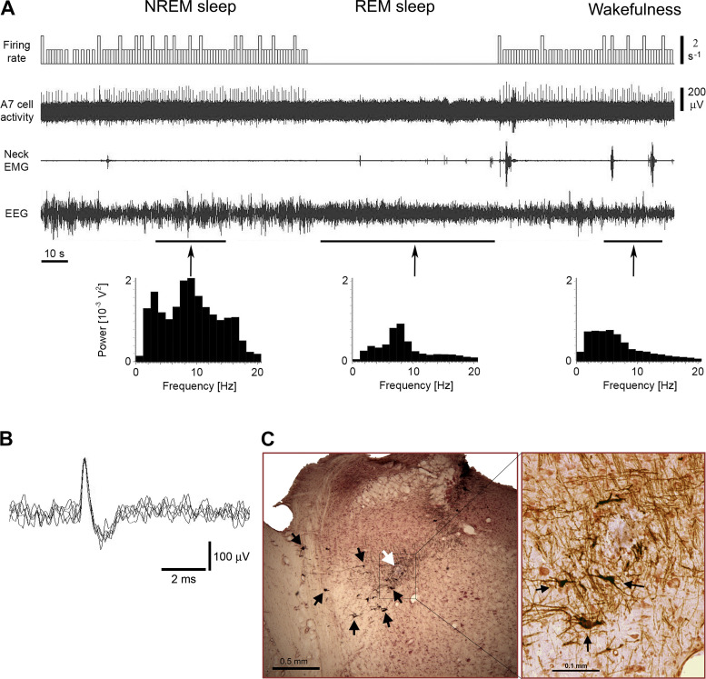
Figure 5.
Extracellular recording of the activity of a putative noradrenergic A7 neuron. A: the A7 neuron discharged regularly with typically low firing rate during NREM sleep (average firing rate 1.04 s−1, CV 0.426) and wakefulness (average firing rate 1.16 s−1, CV 0.17), and was silent during REM sleep. The trace “Firing rate” shows bars that reflect the number of spikes within equal time intervals (in this case, 1 s) that are not synchronized with the state of animal. Power spectra of EEG that were calculated during time periods marked by arrows helped to properly detect behavioral states of the animal. Note the appearance of the theta rhythm in the EEG power spectrum during REM sleep in 7–8 Hz bands. B: superimposed five traces of the A7 neuron action potentials. C: a coronal pontine section stained by immunohistochemistry for TH and neutral red shows the recording site of this A7 neuron (white arrow) within the “loose” part of the A7 nucleus. Black arrows point to some TH-positive noradrenergic A7 neurons with their dendrites within the nucleus. The site was marked by iontophoretically applied Pontamine sky blue from the recording electrode after the end of recording. The black box indicates an area that is expanded and shown to the right. The expanded image shows additional TH-positive A7 neurons nearby the recording site. CV, coefficient of variability; NREM, non-rapid-eye-movement; REM, rapid-eye-movement; TH, tyrosine hydroxylase.

