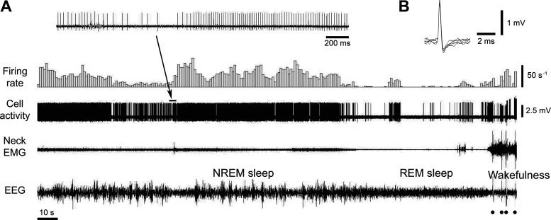
Figure 12.
The activity of a trigeminal motoneuron that was recorded extracellularly during consecutive states of NREM sleep, REM sleep, and wakefulness. Typically, trigeminal motoneurons had relatively high amplitude “short” biphasic action potentials (B, superimposed five traces of action potential). They discharged with a varying frequency (up to 50 s−1) during NREM sleep (average firing rate of this motoneuron during NREM sleep was 19.4 s−1, CV 1.18). During REM sleep-induced muscle atonia, this motoneuron exhibited long periods of silence interrupted by sporadically appeared phasic discharges (average firing rate 2.65 s−1, CV 3.26). During wakefulness, its activity had mostly phasic pattern (average firing rate 21.2 s−1, CV 3.33). Filled circles indicate animal movement artifacts. The inset on A shows the motoneuron activity at expanded time scale. CV, coefficient of variability; NREM, non-rapid-eye-movement; REM, rapid-eye-movement.

