. 2021 Dec 28;2(4):343–360. doi: 10.12336/biomatertransl.2021.04.008

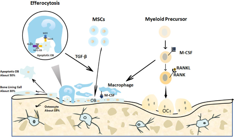
Figure 3. Endogenous cellular changes after bone fracture. When a bone is fractured, the MSCs migrate to the bone defect area and differentiate into osteoblasts to form and remodel the bone matrix. In the end, approximately 15% of the osteoblasts become embedded in the bone matrix as osteocytes, 30% of the osteoblasts become quiescent bone lining cells, and the remaining 40-70% of the osteoblasts are likely to undergo death by apoptosis. The apoptotic osteoblasts expressing certain signals are efficiently cleared by macrophages in a process called efferocytosis. This process is initiated by the expression of the apoptotic signals on osteoblasts and is activated by the binding of linking proteins, including MFG-E8 or Gas6, and macrophage proteins, such as αvβ3 or Mer. The efferocytosis-induced production of specific proteins, such as TGF-β, may promote continuous bone modelling by recruiting osteoblasts from progenitor cells.29 Gas6: growth arrest-specific 6; M-CSF: macrophage colony-stimulating factor; MER (tk): receptor tyrosine kinase MerTK; MFG-8: milk fat globule-epidermal growth factor 8; MSCs: mesenchymal stromal cells; OB: osteoblasts; OC: osteoclasts; RANK: receptor activator of nuclear factor-κB; RANKL: receptor activator of nuclear factor-κB ligand; TGF-β: transforming growth factor β; αvβ3: alpha-V beta-3 integrin.

Figure 3