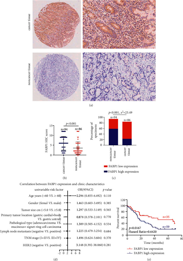
Figure 6.

The expression and clinical significance of FABP1 in gastric cancer. (a, b) Tissue microarray (TMA) analysis by IHC staining showed that a high expression of FABP1 was observed in gastric cancer tissues. (c) High and low expression rates of FABP1 in gastric cancer tissue. (d) Correlations of FABP1 expression levels in gastric cancer tissues and clinicopathological features. (e) Kaplan–Meier analysis showed that gastric cancer patients with upregulation of FABP1 were positively correlated with worse prognosis and shorter overall survival.
