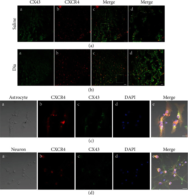
Figure 9.

Confocal images show coexpression of CXCR4 and CX43 in the spinal dorsal horn of rats after STZ-induced DNP at 5 weeks of diabetes and in saline control rats (a, b), and in primary culture astrocytes (c) and neurons (d). (a) and (b) co-expression of CX43 and CXCR4 of spinal dorsal cord were shown in confocal images. (a) (A, B) and (b) (A, B) were double-immunostaining for CX43 (green, A) and CXCR4 (red, B). (a) (C) and (b) (C) were merged images A and B (original magnification: 200×, scale bar 20 μm). Merged and enlarged images were shown in (a) (D) and (b) (D) (original magnification: 400×, scale bar 10 μm, n = 6/group). (c) (A) and (d) (A) were bright field images of astrocytes and neurons, respectively. (c, d) Double-immunostaining for CXCR4 (red, B), CX43 (green, C), DAPI (blue, D). (c) (E) and (d) (E) were merged images A, B, C, and D (original magnification: 400×, scale bar 20 μm, n = 6/group).
