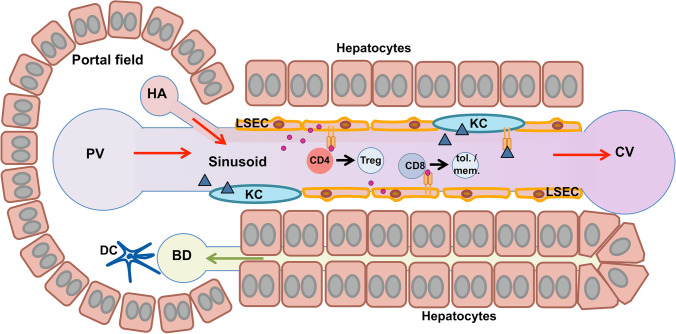
Fig. 2.
Hepatic antigen-presenting cells in anatomical context. Blood flow (red arrows) enters the liver sinusoids through the portal vein (PV) and the hepatic artery (HA) and leaves through the central vein (CV). The hepatic sinusoids are lined by the liver sinusoidal endothelial cells (LSECs), which are scavenger cells clearing the blood from small particles and macromolecules by receptor-mediated endocytosis. LSECs present collected antigens to lymphocytes, producing a state of immune tolerance. CD4 effector T cells (CD4) can be transformed into regulatory T cells (Treg) through TGF-beta signals. CD8 T cells (CD8) can become tolerant or memory T cells (tol./mem.). Kupffer cells (KCs) reside in the lumen of the hepatic sinusoids and facilitate the removal of larger blood-borne particles by phagocytosis. They also present collected antigens to lymphocytes, producing tolerance. Dendritic cells (DCs) predominantly locate in the portal fields, and often close to bile ducts (BD), where they function as sentinels guarding the integrity of the biliary epithelium. DCs are antigen-presenting cells, producing tolerance in homoeostatic conditions, but readily promote inflammation upon sensing of cell damage or infection. As LSECs and KCs are the predominant cells in sinusoidal blood, it is easier to target those than liver DCs with vectors or carriers for antigen-specific immunotherapy

