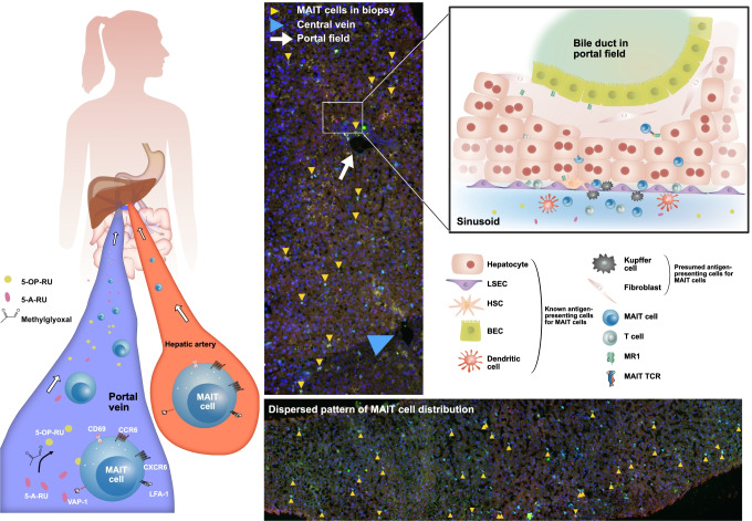
Fig. 3.
MAIT cell homing and distribution in healthy human liver tissue. Left panel: homing of MAIT cells to the liver via the portal vein and the hepatic artery. Examples of homing receptors and integrins (CD69, CXCR6, CCR6, LFA-1, VAP-1) are depicted on a MAIT cell. Gut bacteria-derived MAIT cell stimulatory metabolites are entering the liver via the portal vein. IF staining of tissue sections from liver biopsies without histopathological abnormalities: co-localization of CD3, TCR Vα7.2, and IL18-Rα identifies MAIT cells (yellow arrowheads; methods as in reference [7]). Right panel: cellular interactions of MAIT cells in the liver. Various liver cells including hepatocytes, biliary epithelial cells (BECs), hepatic stellate cells (HSCs), and liver sinusoidal endothelial cells (LSECs) activate MAIT cells in an MR1-dependent manner

