
Figure 3. Cortical lesion morphology is heterogeneous.
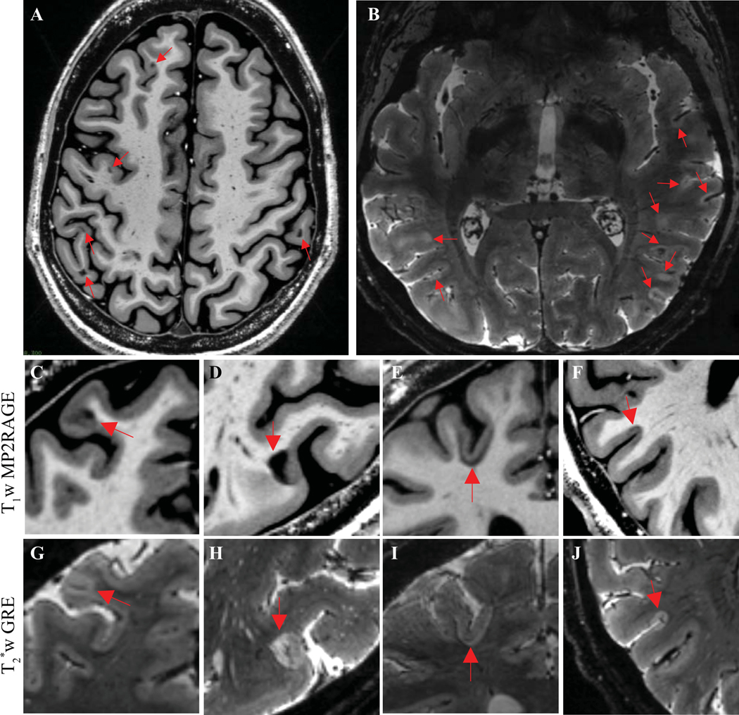
(A) Example of an individual with many juxtacortical lesions but few other white matter or cortical lesions, as seen on a representative T1w MP2RAGE image. (B) Example of an individual with many subpial lesions but few other cortical or white matter lesions, as seen on a representative T2*w image. (C, G) Example of a leukocortical lesion with similar signal change in the cortex and white matter portions of the lesion. (D, H) Example of a leukocortical lesion with marked signal change in the white matter portion of the lesion but more subtle signal change in the cortex, especially on T1w images. (E, I) Example of a lesion involving the juxtacortical U-fibers but relatively sparing the cortex. (F, J) Example of a subpial lesion that stops abruptly at the cortex-white matter border.
T1w MP2RAGE: T1 weighted magnetization prepared 2 rapid gradient echo. T2*w GRE: T2* weighted gradient-recalled echo.