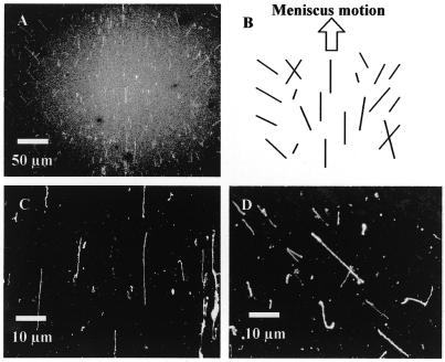
Figure 3.
Straightened λ phage DNAs, combed by meniscus motion control in a <40% humidity environment. These images were captured by a fluorescence microscope utilizing a high sensitivity cooled CCD camera. (A) Overview of a trace of a droplet moved on a coverslip, observed through a 20× objective lens. (B) Schematic of the trend of straightened DNAs in the trace, illustrated on the basis of the direction of meniscus motion and the center line of the trace. (C) Detail of straightened DNAs, observed in the center area of the trace and magnified with a 100× objective lens. (D) Slanted, straightened DNAs seen in the right of the trace.

