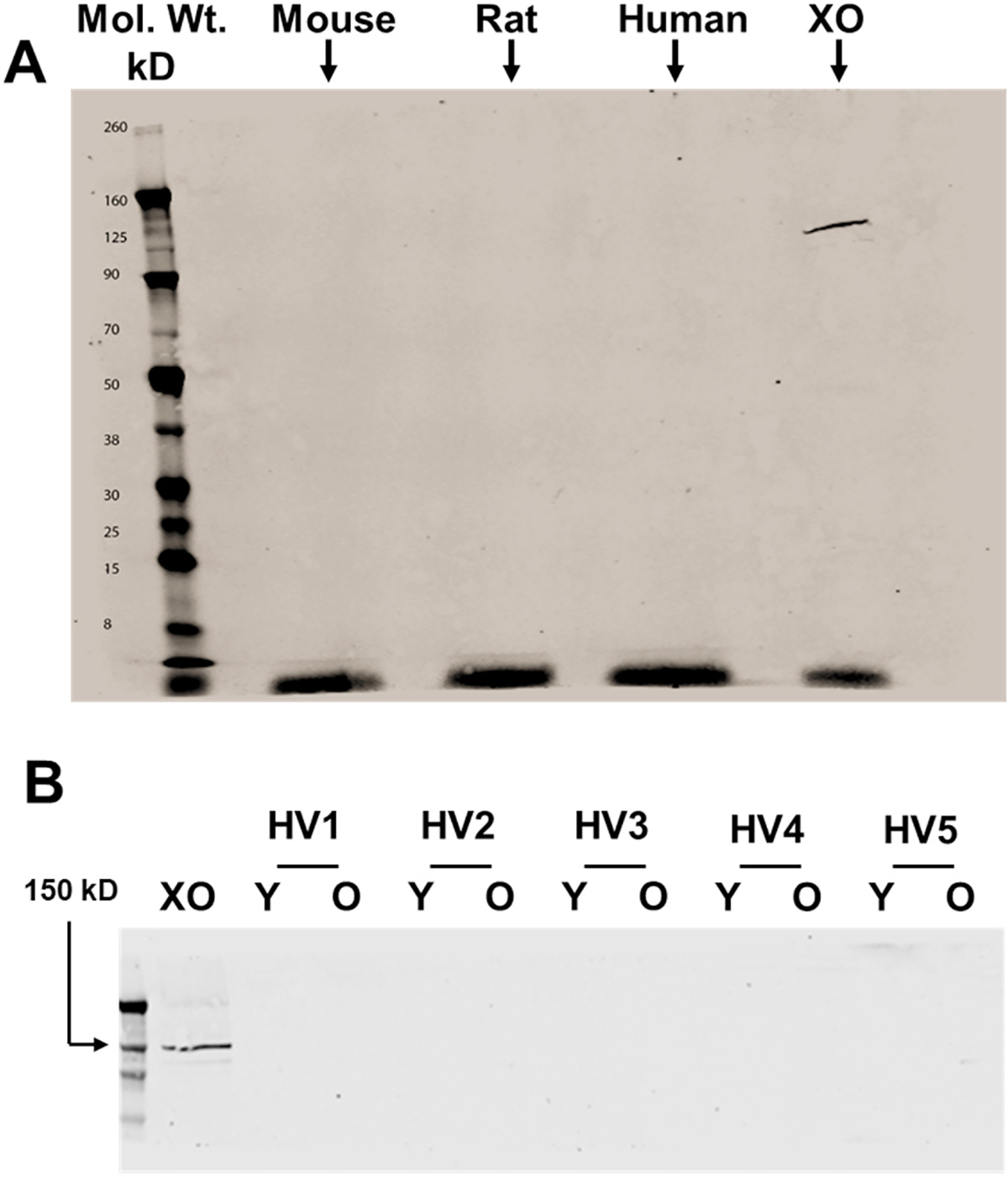
Fig. 3.

Assessment of RBC-associated XOR protein abundance and impact of cellular age. A) Western blot analysis was performed on human, mouse, and rat RBC homogenates using a monoclonal anti-XOR antibody (Santa Cruz) as described in the methods. Purified XO was used as a positive control and electrophoresed using 0.5 μg total protein. A total of 30 μg of protein was electrophoresed for each homogenate (human, mouse, and rat). Shown is a representative of 3 independent blots representing a total of 3 mice, 3 rats and 3 humans. B) Western blot analysis was performed on five different human RBC homogenates, either young or old, using the same mouse monoclonal anti-XOR antibody as described above. Purified XO was used as a positive control and electrophoresed using 0.5 μg total protein. (HV = healthy volunteer/Y = young/O = old).
