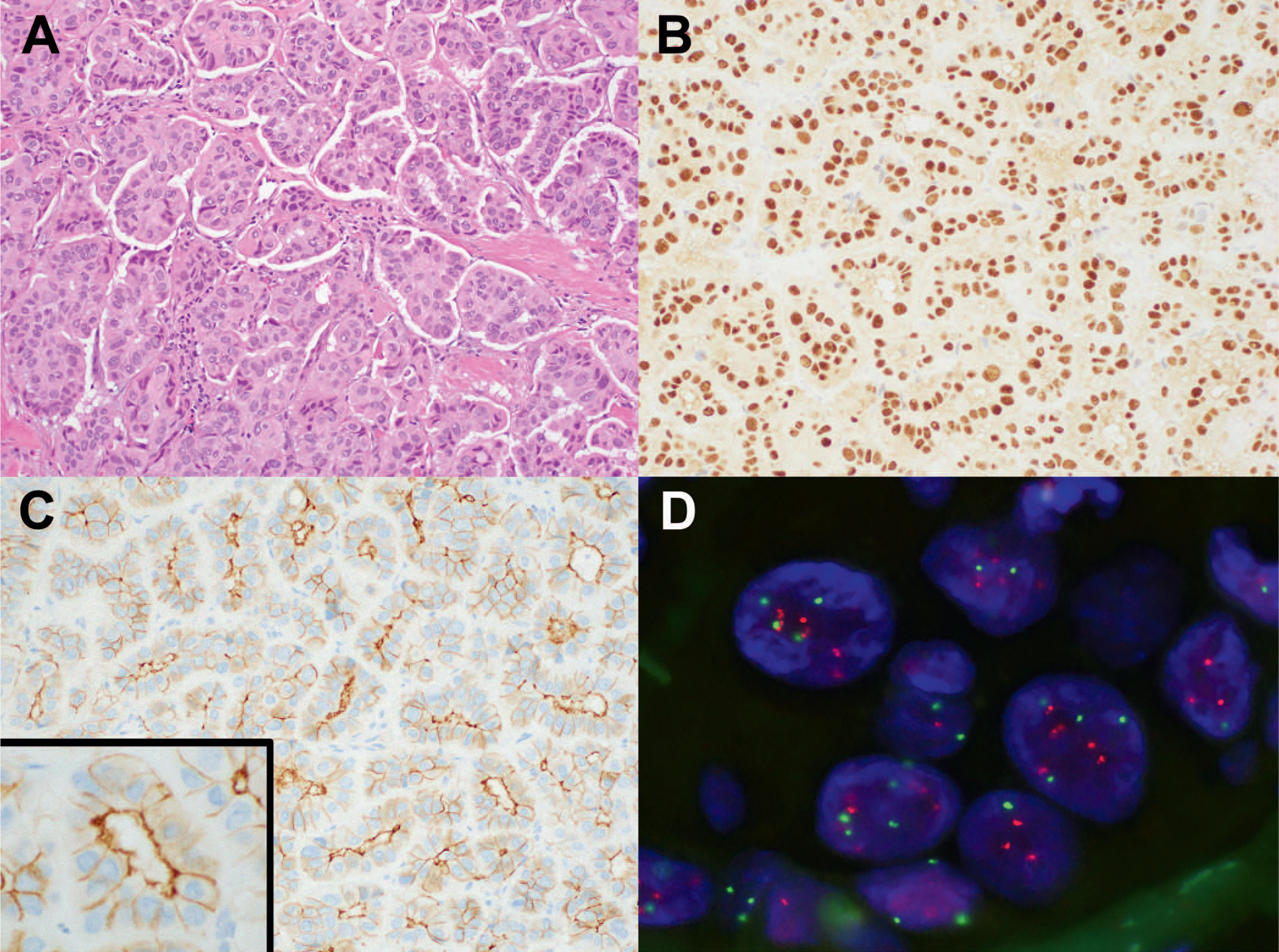
Figure 1.

A, Invasive ductal carcinoma with micropapillary features in a core biopsy. B, ER is diffusely positive with strong nuclear staining. PR (not shown) stained approximately 20% of tumor cells with moderate intensity. C, HER2 immunohistochemistry shows 2+ staining with a “basolateral” pattern. D, HER2 amplification determined by FISH with HER2/CEP17 ratio of 2.2 and 5.2 HER2 signals per cell (hematoxylin-eosin, original magnification ×20 [A]); original magnification ×20 [B]; HER2 immunohistochemistry, original magnifications ×20 [C] and ×40 [inset C]). Abbreviations: ER, estrogen receptor; FISH, fluorescence in situ hybridization; PR, progesterone receptor.
