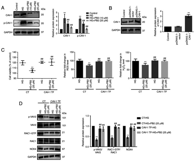
Figure 5.
Effect of CAV-1 overexpression on the cytoprotective effect of PB2 against oxidative stress in Mes13 cells. (A) Cells were treated with PB2 (10 and 25 µM) under HG conditions (25 mM) for 12 h, and the expression levels of CAV-1 and p-CAV-1 were assessed by western blot analysis and statistically analyzed. **P<0.01 vs. control, ##P<0.01 vs. HG. (B) Cells were transfected with pcDNA3.1-vector (negative control) or pcDNA3.1-CAV-1 for 48 h, and the expression levels of CAV-1 were assessed by western blot analysis and statistically analyzed. **P<0.01 vs. control. (C) Cells with or without pcDNA3.1-CAV-1 transfection were treated with PB2 (25 µM) under HG conditions (25 mM) for 12 h. Cell proliferation was assessed by MTT assay. ROS generation was assessed using a ROS assay kit and H2O2 generation was assessed using a H2O2 detection kit, and the results were statistically analyzed. **P<0.01 vs. CT-HG; ##P<0.01 vs. CT-HG + PB2 (25 µM). (D) Cells transfected with or without pcDNA3.1-CAV-1 were treated with PB2 (25 µM) under HG conditions (25 mM) for 12 h. The expression levels of redoxosome-related proteins were assessed by western blot analysis and statistically analyzed. **P<0.01 vs. CT-HG; ##P<0.01 vs. CT-HG + PB2 (25 µM). All data are presented as the mean ± SD of three independent experiments. PB2, procyanidin B2; HG, high glucose; CAV-1, caveolin-1; p-, phosphorylated; NOX4, NADPH oxidase 4; CT, control; TF, CAV-1 transfection.

