
FIGURE 2.
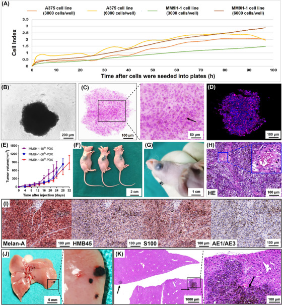
Spheroid formation and tumorigenicity of MM9H‐1 cells and establishment of mucosal melanoma PDX model. (A) Proliferation of MM9H‐1 and A375 cells was monitored by xCELLigence RTCA system. (B‐D) Sphere‐forming assay. (B) Phase contrast image of a sphere from MM9H‐1 cells in 96‐well plate. (C) HE staining shows that MM9H‐1 cells aggregated into regular spheres after 4 days of suspension culture, and the arrow indicates melanin pigmentation in cells. (D) Immunohistochemistry analysis of the sphere shows positive staining for a triple‐antibody cocktail against cytoplasmic HMB45, Melan‐A, and Tyrosinase (red). Hochest was used to stain nucleus (blue). (E‐H) Tumor formation in immunodeficient mice. (E) Tumor growth curves of the subcutaneous tumors formed by the 10th, 50th and 90th passage MM9H‐1 cells (n = 3). Tumor volume was analyzed with Student's t test. Image of tumor formation of MM9H‐1 cells in the subcutaneous (F, 10th passage cells) and orthotopic regions (G, 90th passage cells). (H) H&E staining of the MM9H‐1‐xenografted tumor in subcutaneous regions, with vessel formation indicated by the yellow box. (I) Immunohistochemistry analysis of MM9H‐1‐xenografted tumor shows positive staining for Melan‐A, HMB45, S100 and negative for AE1/AE3. (J) Typical gross appearance of liver metastasis in nude mice injected intrasplenically with MM9H‐1 cells. (K) HE staining for the liver metastasis. The black arrows indicate the metastatic foci with extensive melanin deposition. Abbreviations: HE, hematoxylin and eosin; HMB45, human melanoma black 45