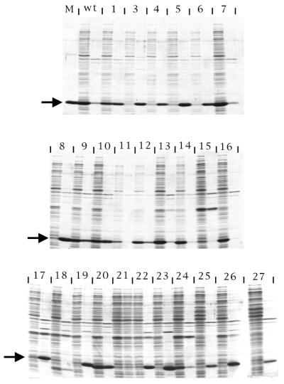
Figure 4.
SDS gel electrophoresis of the soluble (left member of each pair of lanes) and the insoluble (right member of each pair) fractions of lysates produced from cells containing the indicated mutant coat sequences. The lane labeled M contains protein from purified MS2 virus and an arrow to the left of each panel locates the position at which coat protein migrates. WT means wild type. Numbers correspond to the m1 to m27 mutants shown in Figures 2 and 3.

