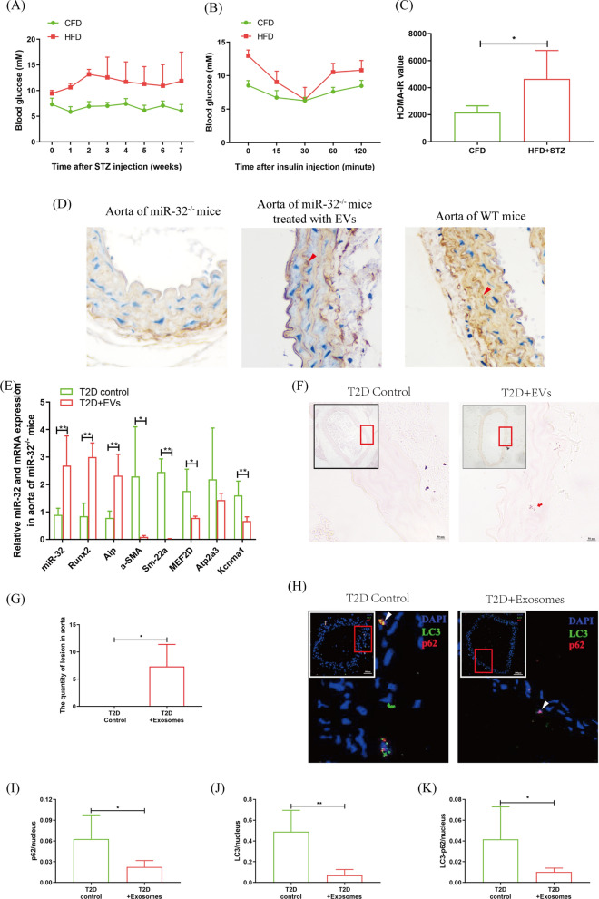
Fig. 6.
In vivo analysis of macrophage EVs miR-32 promotes aorta calcification by inhibited autophagy. Analysis of VSMC autophagy and osteogenic differentiation in the aorta of miR-32−/− T2D mice after treated with macrophage EVs. A Blood glucose analysis. B IPITT analysis. C HOMA-IR analysis. D Immunohistochemisty analyzed miR-32 expression in aorta of miR-32−/− mice, and WT mice, and miR-32−/− injection EVs. C qRT-PCR analyzed aorta osteogenic differentiation. F, G alizarin red staining analyzed aorta calcification after macrophage EVs injection (F), and statistical analysis (G). H–K Immunofluorescence analyzed p62 and LC3 expressions in aorta (H), and statistical analyzed the puncta of p62 (I), LC3 (J), and p62-LC3 (K). 3 per 5 mice in different groups were used in the detection. *p < 0.05 and **p < 0.01. Data was shown as means ± s.e.m

