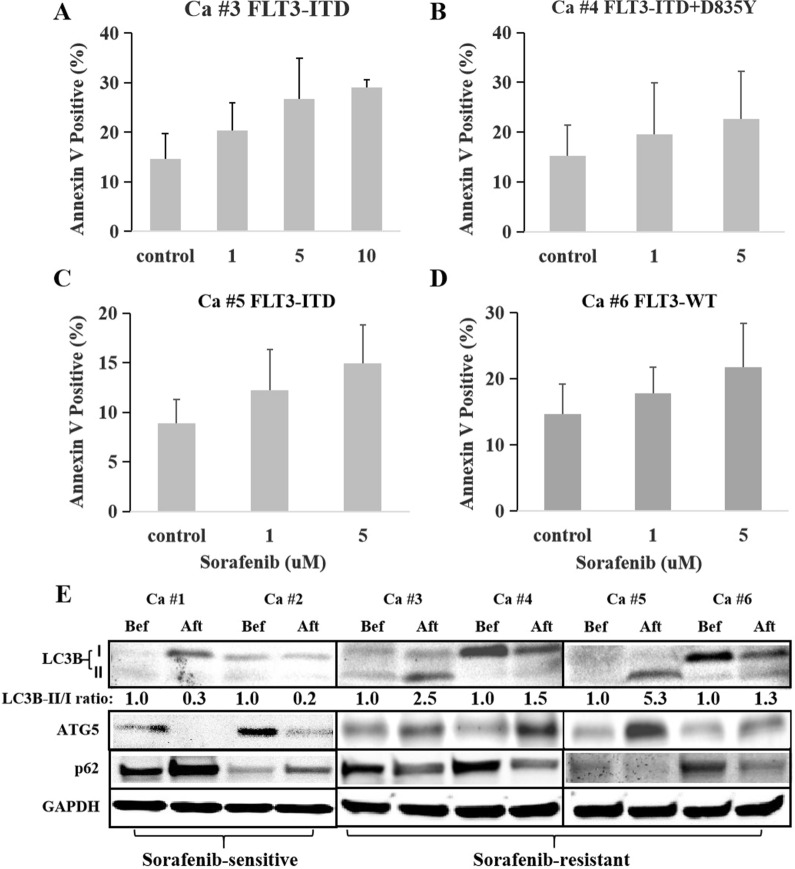
Fig. 1.
Sorafenib-resistant primary AML cells showed high expression of autophagy. A, C Primary AML cells with FLT3-ITD mutation from the patients (case #3 and #5) who relapsed on the maintenance sorafenib therapy were resistant to sorafenib. B Primary AML cells with FLT3-ITD + D835Y mutation from the patient (case #4) who relapsed on the maintenance sorafenib therapy were resistant to sorafenib. D Primary AML cells with FLT3-WT from the patient (case #6) who relapsed on the maintenance sorafenib therapy were resistant to sorafenib. E Sorafenib-sensitive primary AML cells from the patients with FLT3-ITD mutation who were continued complete response (CCR) during sorafenib therapy showed decreasing expression of LC3B-II after vs. before the treatment of sorafenib, showing as LC3B-II/I ratio lower than 1 (0.3 in case #1 and 0.2 in case #2). In contrast to the sorafenib-sensitive cells, sorafenib-resistant AML cells from the patients with FLT3-ITD mutation who relapsed on maintenance sorafenib therapy showed increasing expression of LC3B-II after relapse as compared with before the treatment, presenting as LC3B-II/I ratio higher than 1 (2.5 in case #3, 1.5 in case #4, 5.3 in case #5 and 1.3 in case #6). In accordance with the change of LC3B-II/I ratio, the expression of ATG5 decreased, and p62 accumulated in sorafenib-sensitive cells; while just being opposite, ATG5 increased, and p62 degraded in sorafenib-resistant cells after vs. before the treatment of sorafenib

