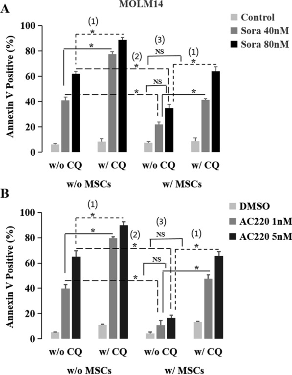
Fig. 7.

Inhibition of autophagy enhanced the anti-leukemia effect of FLT3 inhibitors and overcame MSCs-mediated resistance in FLT3-ITD-positive AML cells. A1 Inhibition of autophagy enhanced the anti-leukemia effect of sorafenib. Regardless of MSCs co-culture or not, CQ significantly enhanced the killing effect of sorafenib in MOLM14 cells, showing as: without MSCs co-culture, the apoptosis rate of 41.2% ± 2.5% w/o CQ vs. 77.4% ± 2.1% w/CQ (P = 0.004) at the concentration of 40 nM, 62.0% ± 1.9% vs. 88.7% ± 2.1% (P = 0.005) at 80 nM; With MSCs co-culture, 22.0% ± 2.1% w/o CQ vs. 41.2% ± 1.3% w/CQ (P = 0.008) at 40 nM, 34.8% ± 3.0% vs. 63.9% ± 3.7% (P = 0.013) at 80 nM, respectively. A2 MSCs decreased the anti-leukemia effect of sorafenib. With MSCs co-culture, the killing effect of sorafenib in MOLM14 cells decreased significantly, showing as the apoptosis rate of 22.0% ± 2.1% vs. 41.2% ± 2.5% w/o MSCs (P = 0.014) at 40 nM and 34.8% ± 3.0% vs. 61.9% ± 1.9% (P = 0.008) at 80 nM, respectively. A3 Inhibition of autophagy overcame MSCs-mediated sorafenib resistance. Though co-culture with MSCs, after being dealt with CQ, the killing effect of sorafenib was similar to that w/o MSCs co-culture, showing as the apoptosis rate of 41.2% ± 1.3% vs. 41.2% ± 2.5% w/o CQ w/o MSCs (P > 0.05) at 40 nM and 63.9% ± 3.7% vs. 62.0% ± 1.9% w/o CQ w/o MSCs (P > 0.05).at 80 nM, respectively. B1 Inhibition of autophagy enhanced the anti-leukemia effect of AC220. B2 MSCs decreased the anti-leukemia effect of AC220. B3 Inhibition of autophagy overcame MSCs-mediated AC220 resistance
