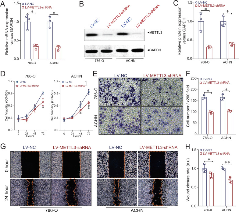
Fig. 2.
Depletion of METTL3 expression in ccRCC cell lines. The stable depletion model of METTL3 in ccRCC cell lines 786-O and ACHN was established by using the RNAi method. A qRT-PCR results showed that the expression of METTL3 at the mRNA level was significantly decreased in LV-METTL3-shRNA compared with LV-NC in both 786-O and ACHN cells (P < 0.05 respectively). B and C Western blot analysis showed that METTL3 expression protein level was significantly decreased in LV-METTL3-shRNA compared with LV-NC in both 786-O and ACHN cells (P < 0.05 respectively). D CCK-8 assay showed that the cell proliferation ability of LV-METTL3-shRNA was significantly inhibited compared with the LV-NC (P < 0.05 in 786-O cells at 72 h, P < 0.05 at 48 h and P < 0.01 at 72 h respectively in ACHN cells). E and F Transwell assay results revealed that the cell invasion ability of LV-METTL3-shRNA was significantly decreased compared with LV-NC (P < 0.05 in 786-O cells, and P < 0.05 in ACHN cells). G and H Wound-healing assay showed that at the time point of 24 h, the relative wound closure rate of LV-METTL3-shRNA was significantly decreased compared with LV-NC (P < 0.05 in 786-O and P < 0.01 in ACHN cells, respectively). Un-paired t test was used as needed. *P < 0.05, **P < 0.01

