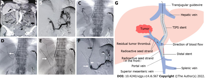
Figure 2.
Representative case. A: Filling defect in the main portal vein (black arrow), suggesting main portal vein tumor thrombosis; B: Most of the intrahepatic branches did not develop under contrast, and several short gastric veins were obviously varicose; C and D: A guidewire was retained in the splenic vein, a catheter was directed into the secondary branch of the right portal vein, and then a radioactive seed strand (white arrow) was implanted; E: Another radioactive seed strand (white arrow) was implanted into another secondary branch of the right portal vein; F: A shunt of transjugular intrahepatic portosystemic shunt (black arrow) was established, a distal stent (short white arrow) was placed, and then a radioactive seed strand (long white arrow) was implanted. Portal venography showed unobstructed blood flow in the shunt and obvious reduction in the varicose veins; G: Schematic diagram. TIPS: Transjugular intrahepatic portosystemic shunt.

