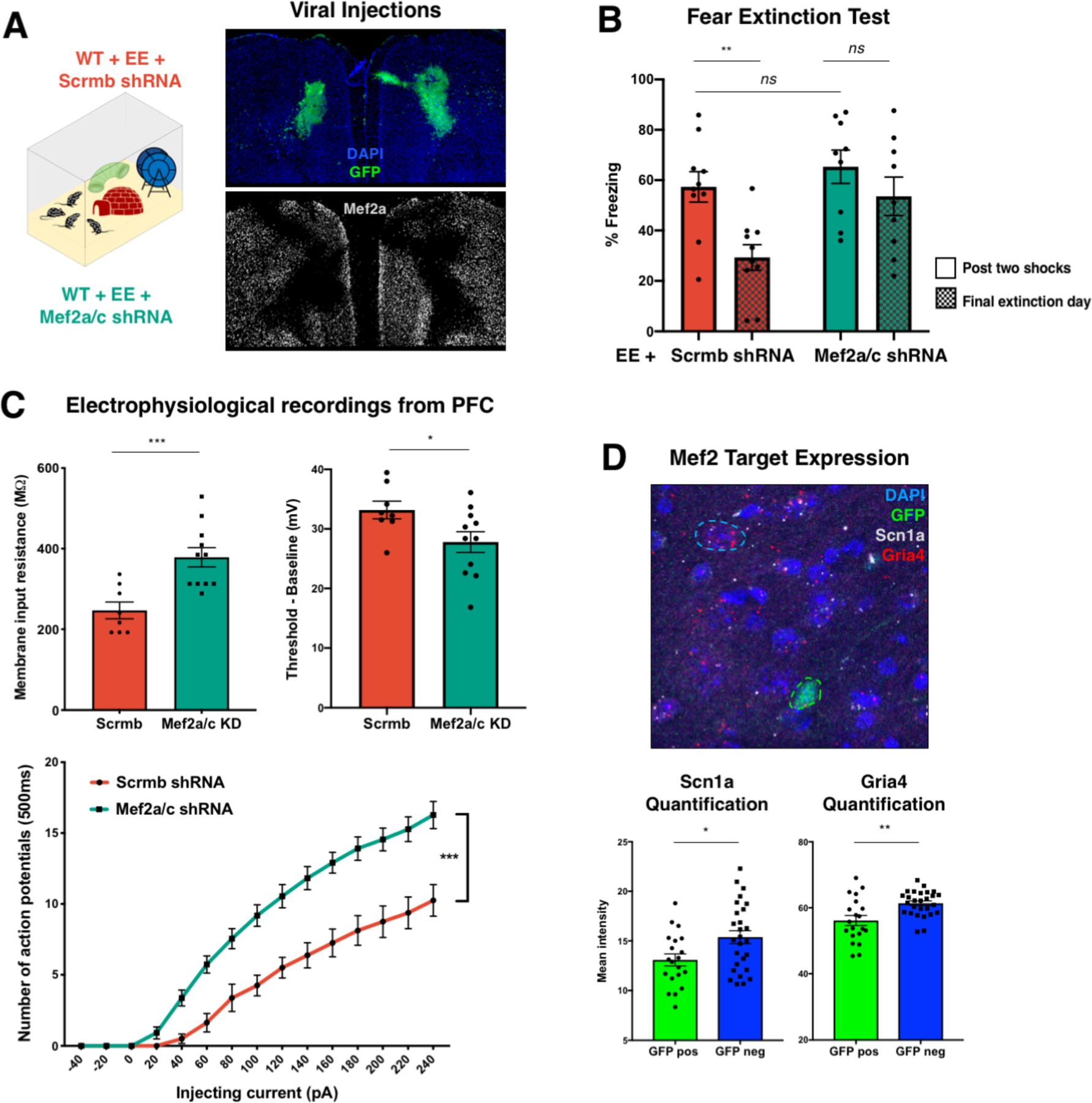
Figure 3 |. Loss of Mef2a/c in the frontal cortex of enriched WT mice impairs cognitive performance and leads to neuronal hyperexcitability.

A, Left: Schematic of experimental paradigm for shRNA knockdown in the context of environmental enrichment. On postnatal day 30, wildtype Swiss Webster mice received bilateral frontal cortex injections of lentiviral vectors containing shRNA cassettes against either a scrambled sequence (Scrmb, red) or Mef2a and Mef2c (Mef2a/c, green). shRNA cassettes also constitutively expressed GFP to aid in determining the knockdown region. Right: Immunohistochemistry staining for DAPI (blue, top), GFP (green, top) and Mef2a (white, bottom), to show representative viral expression and Mef2 knockdown. B, Mef2a/c shRNA-treated mice show significantly impaired fear extinction when compared to Scrmb-treated mice (n = 10 mice per group, unpaired t-test comparing % freezing post 2-shocks and % freezing on last day of extinction training, p=0.3047 for Mef2a/c KD and p=0.0101 for Scrmb). C, Voltage-clamp recordings from pyramidal neurons (KD n = 11 cells from four animals; Ctrl n = 8 cells from 4 animals) in shRNA-expressing regions (identified by GFP fluorescence) revealed that neurons with decreased levels of Mef2a and Mef2c (green) showed increased membrane input resistance (left, unpaired t-test, p-value=0.0010) and a reduced difference between baseline voltage and action potential threshold (right, unpaired t-test, p-value=0.0391). Bottom: Mef2a/c knockdown (green line) was associated with increased hyperexcitability, as measured by number of action potentials in 500 ms for a given injecting current (two-way ANOVA, p-value < 0.0001 for group and interaction). D, Top: RNA in situ hybridization for Scn1a (white) and Gria4 (red) in parallel with immunohistochemistry for GFP (green) in the frontal cortex of animals injected with Mef2a/c shRNA showed decreased levels of Scn1a and Gria4 transcripts in cells expressing GFP along with the shRNA construct (top, representative image). Bottom: Quantification of Scn1a (left; t-test p-value = 0.0168) and Gria4 (right; t-test p-value = 0.0015) intensity in either GFP positive (n=20 from four animals) of GFP negative (n=27 from 4 animals) cells.
