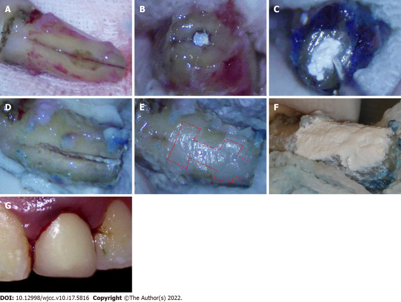
Figure 2.
Operative images. A: A vertical root fracture line after the granulation tissue was removed; B: Excision of the 3-mm apex; C: Filling of the retrograde canal with iRoot BP Plus after retrograde canal preparation; D: Cleaning and enlargement of the fracture line; E: Filling of the fracture and trapezoidal retention forms (shown by dotted line) with resin; F: Covering of the resin surface with iRoot BP Plus; G: Replantation of the tooth.

