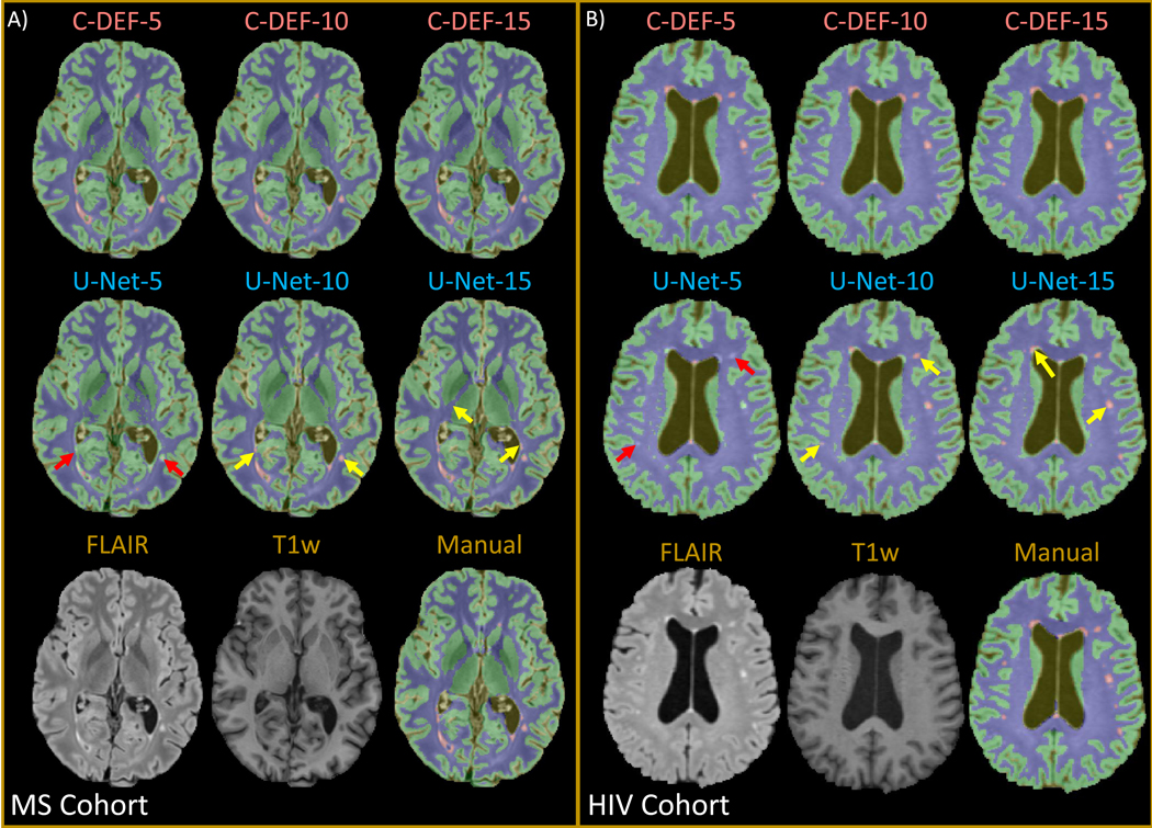
Figure 2: Qualitative assessment of segmentation at various training data sizes:

Output of segmentation performed with varied numbers of training data (5, 10, 15) using C-DEF and U-Net algorithms shown on a representative slice from a participant in (A) (Male, 34 years old) the MS cohort and (B) (Female, 56 years old) the HIV cohort. Bottom row shows pre-processed input scans and the manually drawn mask for reference. Red arrows indicate segmentation errors, while yellow arrows indicate areas of improved segmentation with more training data.
