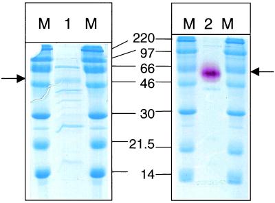
FIG. 1.
PAGE of a protein fraction from conidia of A. sydowi IAM 2544 enriched in frucosyltransferase activity (lanes 1 and 2). Left: the gel was stained with Coomassie brilliant blue. The band with sucrolytic activity is marked by an arrow. Right: activity stain with 1,2,3 triphenyltetrazolium chloride. M, marker lane. The numbers between the two gels give the molecular mass of the marker proteins in kDa.

