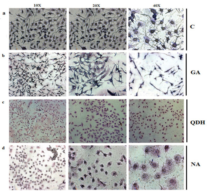
Figure 5.
Giemsa Staining of a) Untreated Cells, (b) GA, (c) QDH and (d) NA. SiHa cells were cultured in 6 well plate and treated with IC50 of respective compounds. Images were captured by inverted microscope after 48hrs. of treatment with different magnification that were 10X, 20X and 40X from left to right

