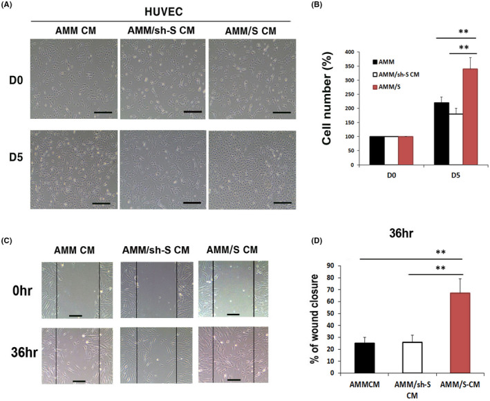
Figure 2.

Cell proliferation, migration assays. (A) Representative photograph of HUVEC proliferation after 5 days incubation with CM. (B) Comparison of HUVEC cell proliferation rates after 5 days showed that AMM/S CM significantly improved HUVEC proliferation compared to control AMM or AMM/sh‐S CM. **p < 0.01, n = 4 in each group. (C) Representative photograph of HUVEC migration after incubation with CM. (D) An in vitro wound scrach healing assay indicating that AMM/S CM strongly enhanced cells migration compared with control AMM or AMM/sh‐S CM. **p < 0.01, n = 4 in each group
