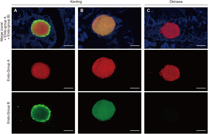
Fig. 3. Visualization of Endozoicomonas phylotypes within individual CAMAs in the tissues of S. pistillata.
Representative confocal images depicting the hybridization of two Endozoicomonadaceae-specific probes (Endo-Group A probe targeting OTUs 2 and 16 and Endo-Group B probe targeting OTUs 5 and 18) labeled with Cy3 (red) and Cy5 (green), respectively. Two different patterns are observed with the Endo-group B probe binding prominent in the CAMA periphery (A): Homogeneous binding of both Endo-group B and Endo-group A is observed in other CAMAs (B) in Kenting samples, while the Endo-Group A probe is exclusively visible in Okinawa samples (C). Scale bars, 20 μm.

