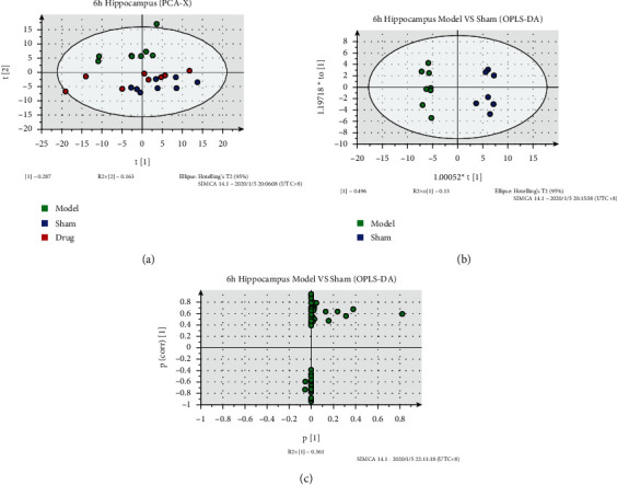
Figure 3.

Metabolic analysis of hippocampus samples at 6 h after cerebral ischemia. (a) PCA-X image of hippocampal samples at 6 h after cerebral ischemia. (b) OPLS-DA of hippocampal samples at 6 h after cerebral ischemia. (c) S-plot[M2] of hippocampal samples at 6 h after cerebral ischemia.
