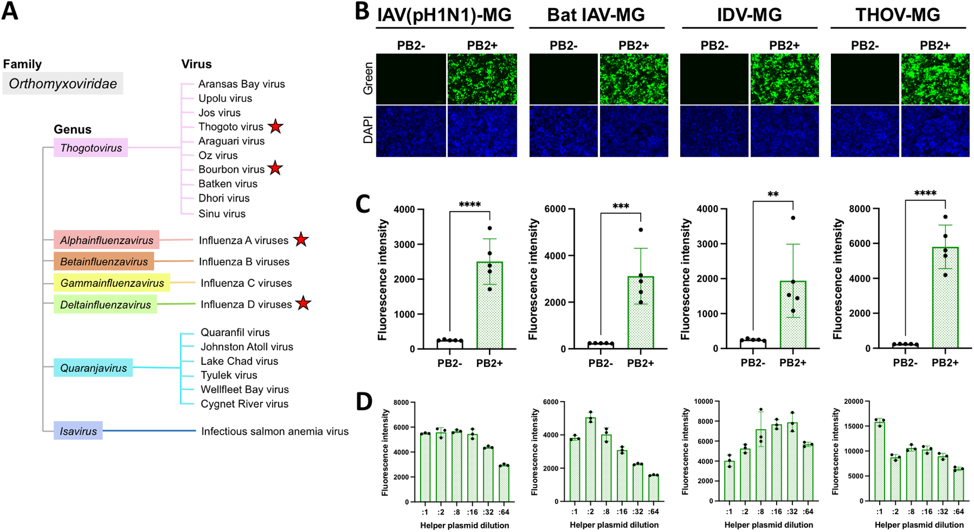
Fig. 6:

Fluorescence-based orthomyxovirus MG systems in HEK293T/17 cells. A) Classification of viruses belonging to the family Orthomyxoviridae. The names of viruses which have been isolated are shown. MG systems were generated for viruses labeled with red stars. MG activities are shown by B) fluorescence signals and C) fluorescence intensity with or without supplementation of PB2 helper plasmid at 72 hours post-transfection. For B) and C), HEK293T/17 cells were seeded in 24-well plates and transfected with a MG-ZsGreen plasmid (0.25 μg) together with helper plasmids (0.1 μg each). D) MG activities induced by transfection of serially diluted helper plasmids. HEK293T/17 cells were seeded in 96-well plates and transfected with an IAV (pH1N1) MG-ZsGreen, IDV MG-ZsGreen, and THOV MG-ZsGreen plasmid (125 ng) or bat IAV MG-ZsGreen plasmid (62.5 ng) together with helper plasmids (25ng each as the initial amount which were further diluted for :2, :8, :16, :32, and :64). Data are representative of the average of three independent experiments or triplicate in one experiment. **: p < 0.005, ***: p < 0.0005, ****: p < 0.00005.
