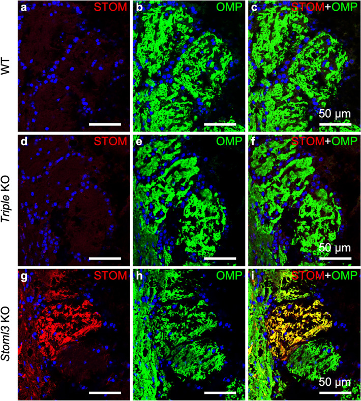
Figure 4.
STOM expression in the olfactory bulb. Confocal micrographs of coronal sections of the OB of WT (a–c), triple KO (d–f), and Stoml3 KO (g–i) mice, double-stained with anti-STOM (red) and anti-OMP (in green) antibodies. In WT mice, STOM is not detected in the OB. Triple KO (d–f) mice do not display any staining with anti-STOM antibodies. In contrast, in Stoml3 KO mice, STOM strongly mis-localizes in the glomeruli (g–i). Nuclei were stained with DAPI (blue).

