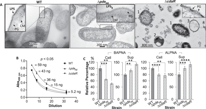
Fig. 5. Effects of defective regulation of the cellular c-di-AMP level on P. gingivalis cell envelope, the immunoreactivity of LPS, and gingipain activities.
A Cells were stained with osmium tetroxide and uranyl acetate and imaged by transmission electron microscopy. Cells of ∆cdaR mutant displayed a significant shape and cell envelope heterogeneity represented by rod- and round-shaped cells with fully or partially intact cell envelopes that are overloaded with OMVs varying in shape and size (1); cells with an intact monolayer of membrane encompassing agglomerated cytoplasmic materials (2) or entirely void round-shaped structures with an intact monolayer of the membrane (3), cells displaying bare peptidoglycan layers (4). B ELISA assays of cell lysates using anti-P. gingivalis LPS monoclonal antibody. The graph represents the mean ± SE of the immunoreactivity of cell lysates (three biological replicates). The standard curve is presented in Supplementary Fig. 10. C Gingipain-dependent proteolytic activities of P. gingivalis strains cells. Graphs represent the mean ± SE (three biological replicates) of the activity of arginine (BAPNA) and lysine (ALPNA) gingipains which were analyzed with a Student’s t test (**P < 0.01; ***P < 0.001; ****P < 0.0001). Scatter plots in Supplementary Fig. 9 display the data distribution. Sup cell-free supernatant, BAPNA N-α-benzoyl-l-arginine-p-nitroanilide, ALPNA N-α-acetyl-l-lysine-p-nitroanilide.

