FIGURE 4.
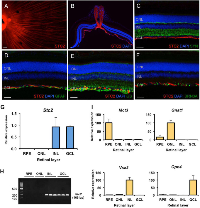
Site-specific detection of STC2 protein and mRNA expression in retinal tissue. (A) Immunostaining for STC2 (red) in a retinal flatmount of a 7 weeks old wild type (129S6) mouse. (B) Immunostaining for STC2 in a retinal section of an 11 weeks old wild type (129S6) mouse. Co-immunostainings for STC2 (red) and SYN (C, green), GFAP (D, green), GS (E, green), and BRN3A (F, green). Nuclei are stained with DAPI (blue). Scale bar: 50 μM. (G) Relative expression of Stc2 mRNA in the RPE (retinal pigment epithelium), ONL (outer nuclear layer), INL (inner nuclear layer), and GCL (ganglion cell layer) that were isolated by laser capture microdissection. (H) The amplified PCR products from experiments shown in panel (G) were run on an agarose gel for visualization. N = 3. (I) Relative expression of layer-specific control genes: Mct3 for RPE, Gnat1 for ONL, Vsx2 for INL, and Opn4 for GCL. Expression was determined by semi-quantitative real-time PCR and normalized to Actb. Shown are mean values ± SD of N = 3.

