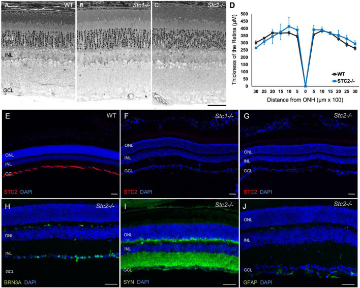
FIGURE 5.
Retinal morphology in the absence of STC1 and STC2. Retinal morphology of wild type (WT, C57BL/6) (A), Stc1–/– (B) and Stc2–/–, (C) mice. (D) Spidergram of the retinal thickness of WT (C57BL/6, black line) and Stc2–/– (blue line) mice; N = 3, ONH: Optic Nerve Head Immunostainings for STC2 (red) in retinal sections of wild type (C57BL/6) (E), Stc1–/– (F), and Stc2–/– (G) mice. Immunostainings for BRN3A (H), SYN (I), and GFAP (J) in retinal sections of Stc2–/– mice. DAPI (blue) was used as nuclear stain. Images were acquired at 200 μm from the optic nerve head. Mice were 6 months (wild type) or 9 months (Stc1–/– and Stc2–/–) of age. Shown are representative images of N = 3 retinas per genotype. Scale bar: 50 μm.

