
Figure 3. Bacterial envelopes and receptors for tailed phages.
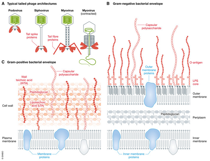
(A) Schematic overview of the three typical tailed phage architectures. Fibre and spike components in the baseplate that interact with bacterial envelope glycans are shown in red. (B) Gram‐negative bacterial envelope. (C) Gram‐positive bacterial envelope with glycan (red) and protein (blue) phage receptors.