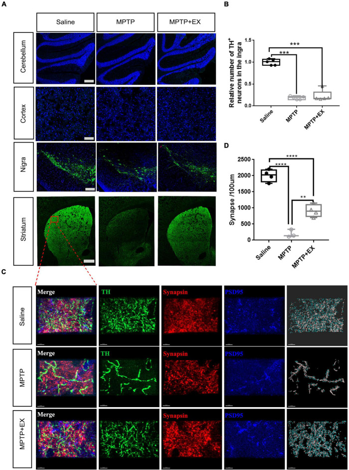
FIGURE 5.
Immunohistochemical staining experiments validate synaptogenesis involved in exercise-induced PD recovery. (A) TH+ staining in the cerebellum (scale bar, 200 μm), cortex (scale bar, 200 μm), SN (scale bar, 200 μm), and striatum (scale bar, 500 μm). (B) Relative number of TH+ cells in the SN for Saline, MPTP, and MPTP + EX groups (n = 5 per group). (C) Immunohistochemical staining of TH+ fibers (green) stained with synaptic markers in striatum (scale bar, 5 μm). Presynaptic terminals and postsynaptic structures were stained by synapsin (Syn, red) and postsynaptic density protein 95 (PSD95, blue), respectively. Synapses were identified by the close proximity of pre- and postsynaptic elements (< 1 μm). A 3D construction of PSD-95 and presynaptic synapsin was performed with “create spots” algorithm in Imaris (scale bar, 5 μm). (D) Quantitation of synapses in the striatum for saline, MPTP, and MPTP + EX groups (n = 3–5 per group). Data are mean ± s.e.m. Statistical analysis was performed using one-way ANOVA followed by Tukey’s post hoc test; **p < 0.01, ***p < 0.001, ****p < 0.0001.

