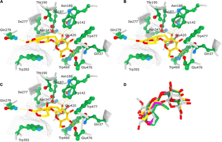
FIGURE 10.
Position of docked ligands in the CeBGlu1 active site. Interactions of docked ligands: gentiopicrin (A), sweroside (B) and swertiamarin (C) with the CeBGlu1 active site residues. Ligands are colored yellow with oxygen shown in red; active site residues are colored green with oxygen shown in red and nitrogen in blue. H-bonds are indicated with yellow dashed lines while the hydrophobic surface encompassing the aglycone is indicated with a silver mesh. (D) Superposition of ligands docked to the CeBGlu1 active: gentiopicrin (yellow), sweroside (pink) and swertiamarin (green) with secologanin (gray) from the experimental structure of glucosidase from Rauvolfia serpentina (PDB: 3U5Y). Superposition of CeBGlu1 with 3U5Y was performed using matchmaker command in ChimeraX using default options.

