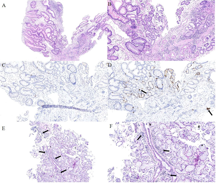
Figure 3.
Histopathology of gastric mucosa and labial salivary gland. (A) Hematoxylin and eosin staining (40×) displays a hyperplastic polyp in the corpus. (B) Hematoxylin and eosin staining (100×) displays the background of the hyperplastic polyp: atrophy, intestinal metaplasia and pseudo-pyloric adenylation in the mucosa. (C)Immunohistochemical staining of gastrin is negative in pseudo-pyloric gland. (D) Staining with anti-chromogranin antibodies (CgA) (100×) depicts dark brown endocrine cells (arrow). (E) Hematoxylin and eosin staining (100×) displays no obvious atrophy in the acinus of the labial salivary gland, but small and numerous foci of lymphocytic aggregation was observed (arrow). (F) The arrow indicates the squeeze of the lymphocytes.

